Stone statues
Select a case study:

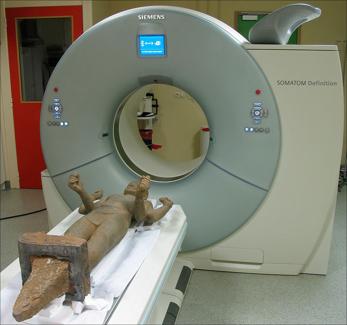
No literature has yet described CT scanning of monolithic sculptures. The reason for this is probably to be found in the apparent incompatibility between the relatively low penetrating power of
the X-rays used in medical CT scanners (compared with industrial CT scanners) and the high density of the stone.
Yet experience shows that medical CT scanners are powerful enough for stone sculptures under 40 cm in diameter. A CT scan is even a valuable source of information on the inner state of the
material, which cannot be established with the other types of scientific analysis, because they mostly study the surface of the sculpture or samples taken from it.
Thus the CT can show whether a sculpture has been constructed from one block or several. If there are several, it can show whether or not they are of the same nature by analyzing their density,
the direction of their veins or sedimentary strata, and the quantity of natural metals they contain.
If repairs have been made, separate elements such as metal rods, drill holes, cement joins and injected resin deep within the stone show up clearly.
Fakes can also be detected, such as a head attached to a body made of a different stone.
Lastly, the CT scan can give information about the outer crust and even find deep-lying causes for surface anomalies. Thus a crack running around the statue does not necessarily mean that the
stone has been broken and glued together again, but may be due to the natural erosion of an oxidized sedimentary stratum.