
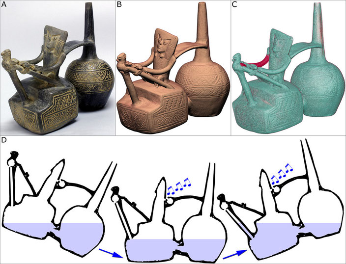
Peruvian Bottle

Beside its medical use, CT scanner can be used to reveal the functions or mechanisms of antique ceramics.
The bottle illustrated above works on the principle that liquid moving through a narrow passageway from one chamber to another displaces air in the second chamber where two whistles have been
enclosed, one in the bird and one on the back of the weaver. When the chambers are approximately half full of liquid, the bottle can be tilted so that the water will push air across the sounding
edge of the whistles and produce a whistling noise. As illustrated on the sagittal reconstructions, the bottle makes a whistling sound each time it is tilted back and forth.
Even if this Chimú bottle (Peru, 900-1,200 A.D.) was primarily CT scanned to check the extent of restoration on the weaver’s right arm and the left wing of the bird sitting on the loom, the
revelation of the enclosed whistles is interesting as it shows how whistles were very precisely fashioned from a hollow sphere of clay with a small hole on one side. A tube of clay was then
positioned adjacent to the outer surface of the sphere so that, after firing, the stream of air would be correctly directed across the opening. As this air crosses the opening, it resonates,
causing a whistling sound. The CT scan exposure of this technical device for forming ceramic whistles highlights only one of the many innovations developed by ancient Peruvian potters over a
period of more than 3,000 years.
Video CT scan: 3D half opaque VRT view
Video CT scan: 3D opaque VRT view
Video CT scan: 3D opaque VRT view with various manipulations
Video CT scan: 3D transparent VRT view
Video CT scan: 2D MPR sagittal slices